Digital slide repository
|
HTML5 viewer NZH viewer |
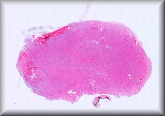
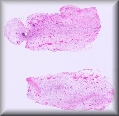
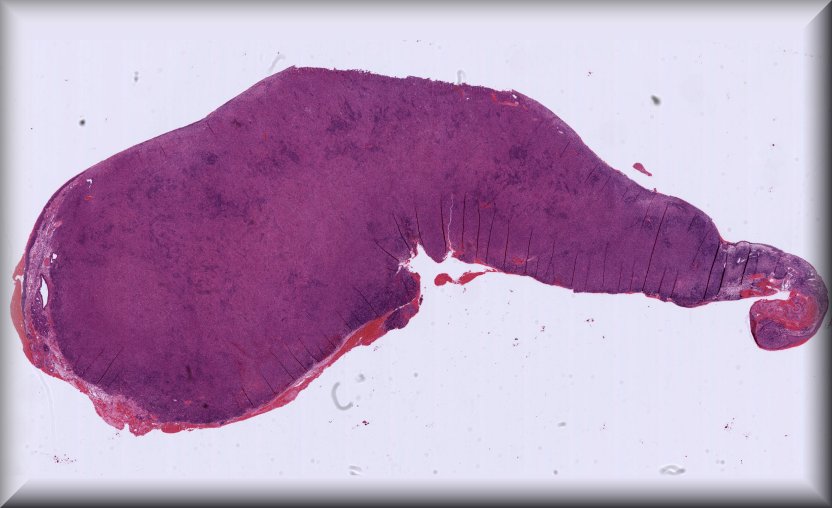
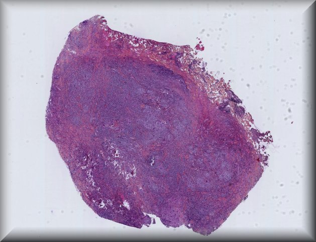
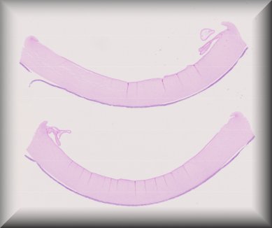
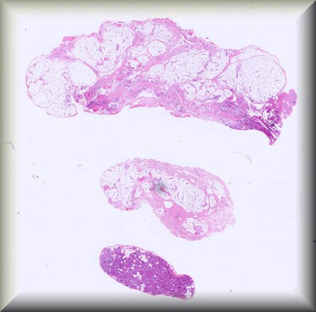
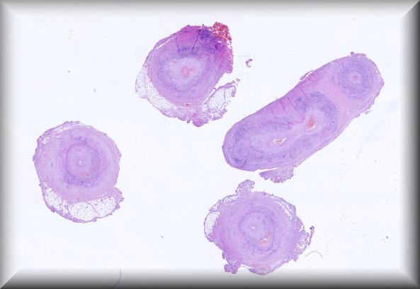
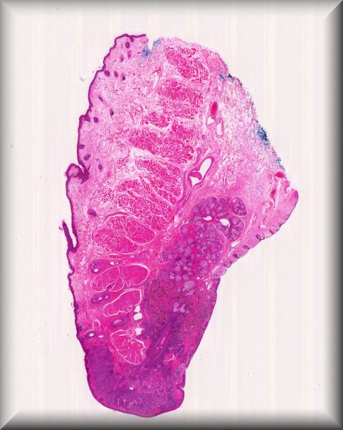
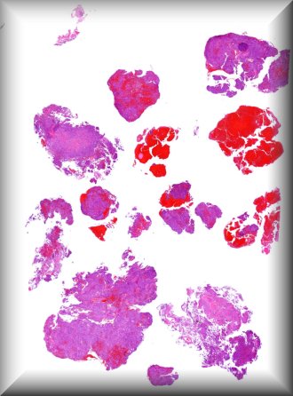
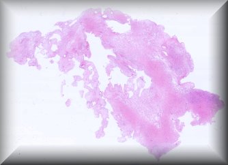
María Antonia Saornil (Valladolid): 19-year-old male patient who, in the last 3 months, had a progressive and extensive protrusion in the temporal sclera of the left eye. The patient also reports noticing loss of nasal visual field. |
||
 |
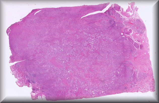
HTML5 viewer NZH viewer |
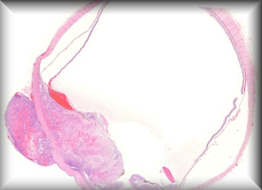
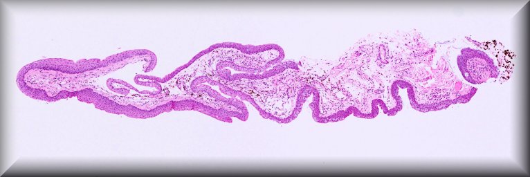
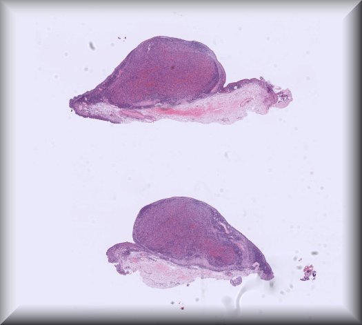
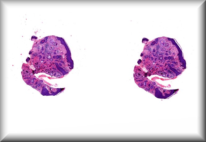
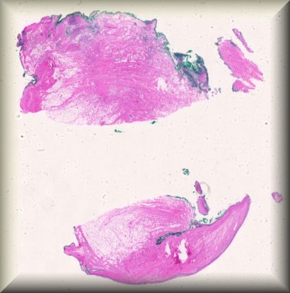
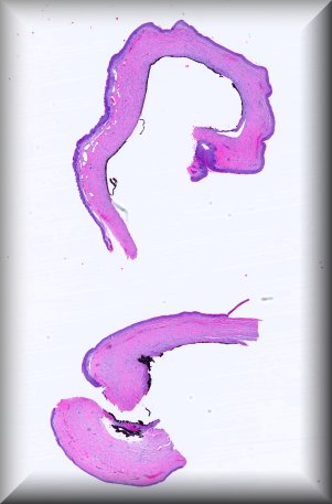
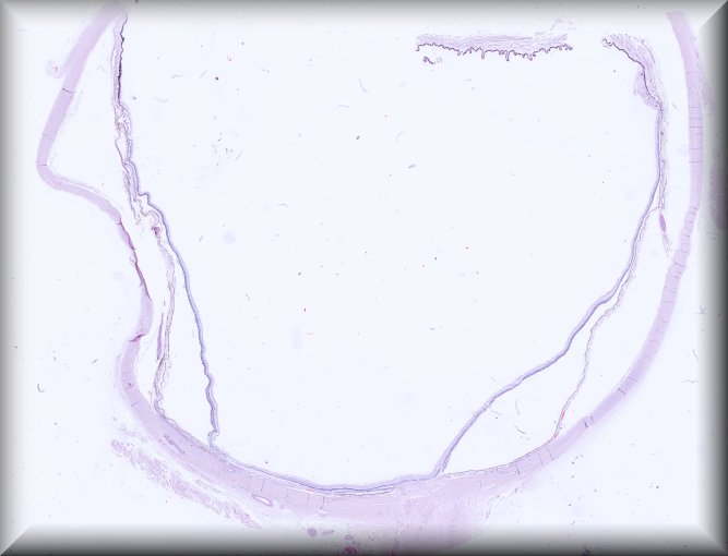
Daniela Süsskind (Tuebingen): Rapidly growing tumor of the conjunctiva in an 86 years old woman presenting as a masquerade syndrome with signs of intraocular inflammation. |
||
 |
HTML5 viewer NZH viewer |
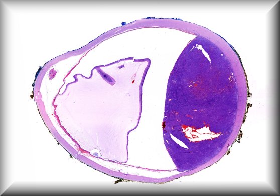
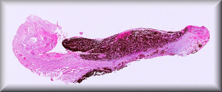
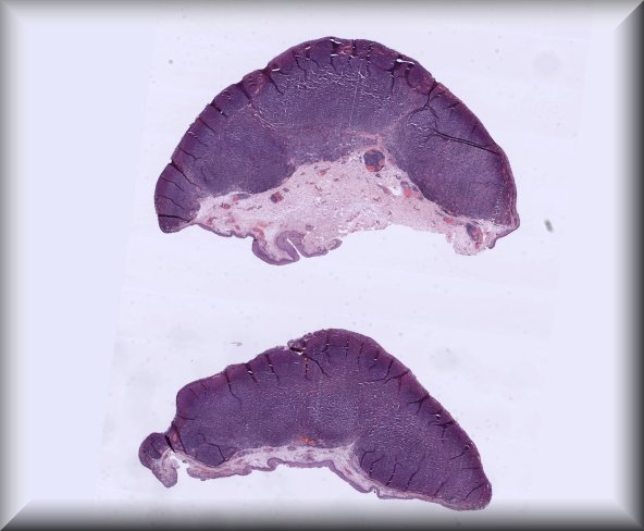
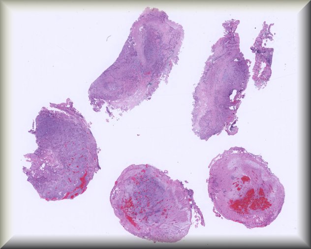
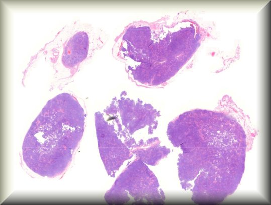
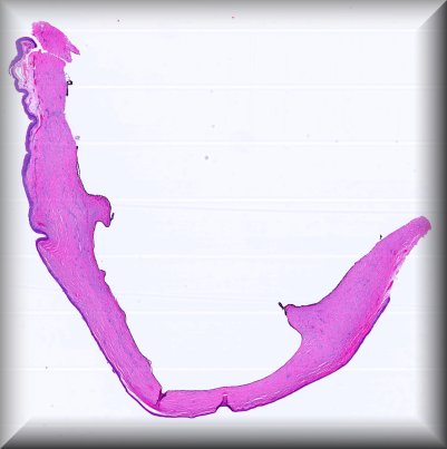
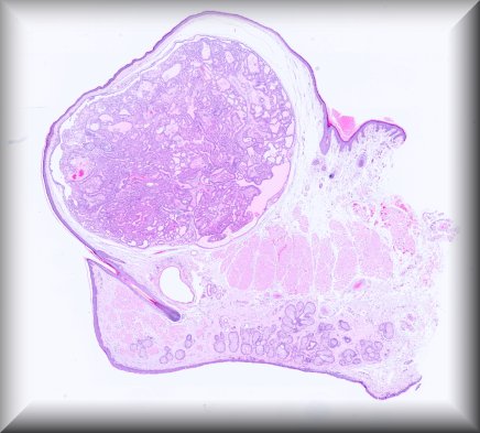
Dietmar Thal (Leuven): Eyeball resection for melanoma in an 83-year old male Alzheimer's disease patient |
||
 |
HTML5 viewer NZH viewer |
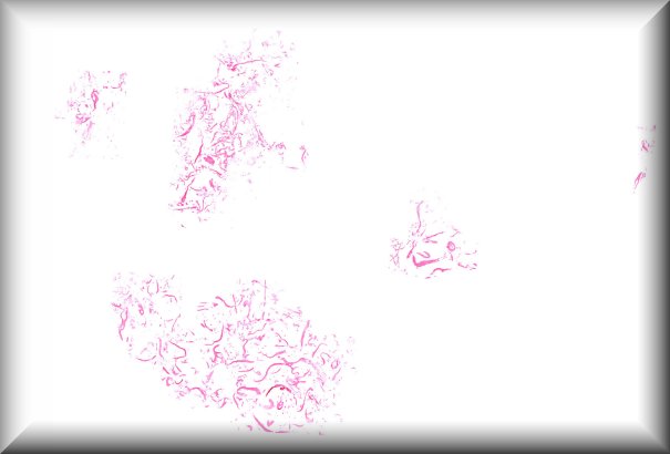
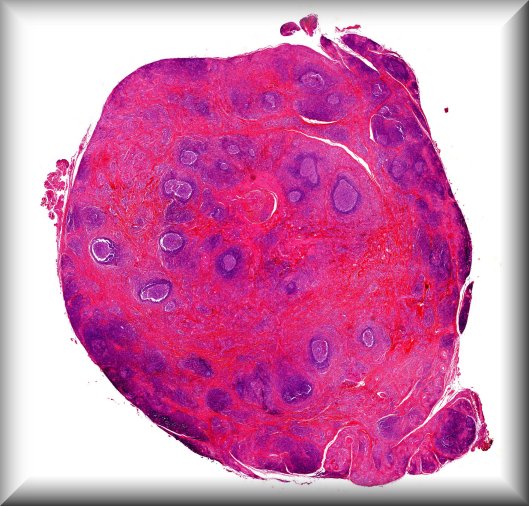
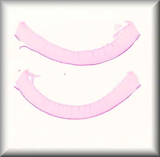
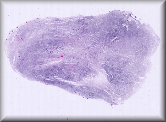
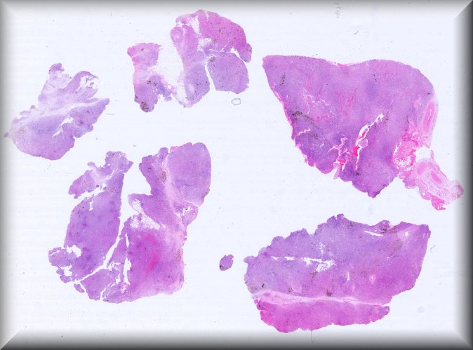
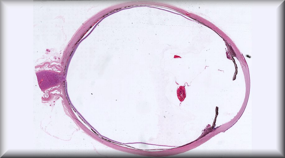
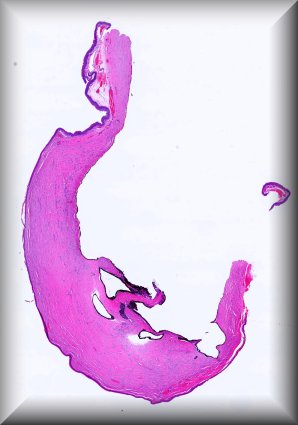
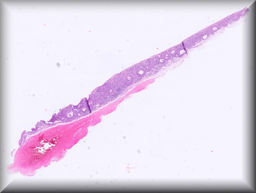
Rita Van Ginderdeuren (Leuven): Vitreous sample in chronic posterior uveitis, unresponsive to steroid treatment |
||
 |
HTML5 viewer NZH viewer |
|||
 |
HTML5 viewer NZH viewer |
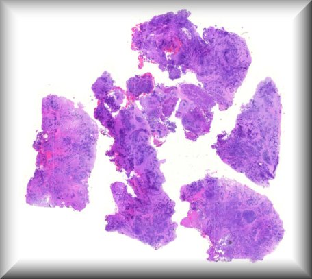
Claudia Auw-Haedrich (Freiburg): Atypical lipomatous tumour of the orbit in a 70-year-old lady [previous biopsy I] [previous biopsy II] |
||
 |
HTML5 viewer NZH viewer |
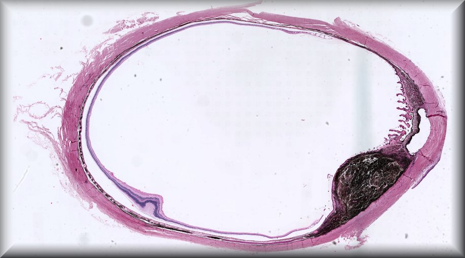
Simone Nuessle (Freiburg): Rapidly growing dark scleral mass. |
||
 |
HTML5 viewer NZH viewer |
|||
 |
HTML5 viewer NZH viewer |
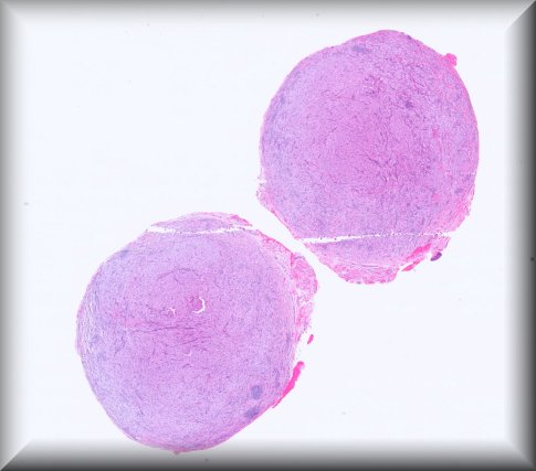
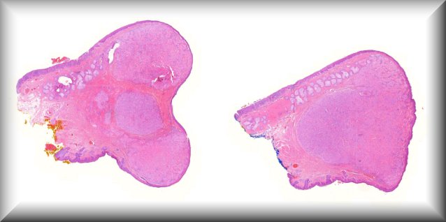
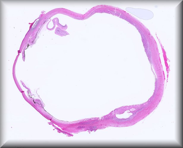
Mª Rosa Bella (Barcelona): 54-year-old woman who presented with a nodule in the orbitonasal angle of the right eye, hard and adherent, slow growing (from 1 cm to 1.5 cm in diameter in 3.5 years), for which a simple resection was performed. A section of the lesion is sent. |
||
 |
HTML5 viewer NZH viewer |
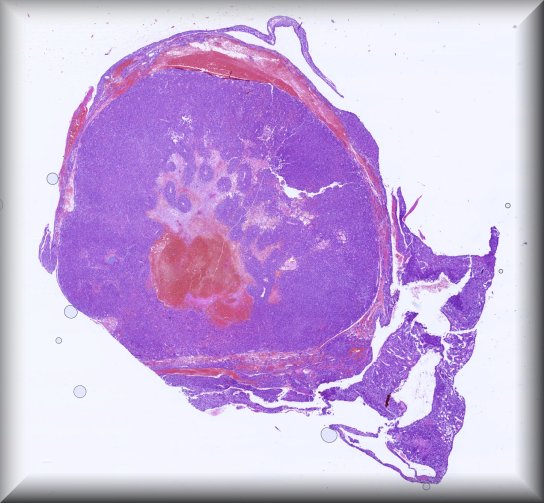
Sarah Coupland (Liverpool): An 83-year old West African female patient presented with a large rapidly-growing right conjunctival tumour. She had a complex background history of bilateral eye disease, including bilateral cicatrizing conjunctival disease of unknown cause leading to bilateral corneal decompensation, penetrating keratoplasty (PK) of the right eye that was complicated by reinfection of right eye PK with pseudomonas infection, as well as left eye non-healing pseudomonas corneal ulceration resulting in left eye enucleation. |
||
 |
HTML5 viewer NZH viewer |
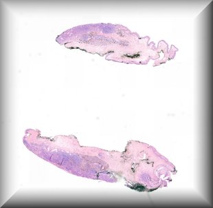
Luciane Dreher Irion (Manchester): 43-year-old female with a long standing right lower eyelid presumed sebaceous cyst. |
||
 |
HTML5 viewer NZH viewer |
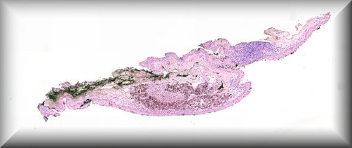
Jolique van Ipenburg (Nijmegen): A thirteen year old girl presents with two conjunctival lesions. The patient reported a traumatic event 2 years earlier, where after she noticed these conjunctival lesions 1 ½ year ago. These lesions seem to become darker over time and people are making remarksfrequently. Clinical examination reveals 2 heavily pigmented mobile highly vascularized lesions located at the bulbar conjunctiva. These lesions do not bleed and are painless. Vision is not impaired. The lesions were excised for both diagnostic and therapeutic reasons. |
||
 |
HTML5 viewer NZH viewer |
|||
 |
HTML5 viewer NZH viewer |
Sandra Lassalle (Nice): 23-year-old man with no comorbidities presenting with an orbital mass that had been evolving for 1 month. The mass was growing rapidly, leading to complete closure of the eye. Antibiotic treatment for suspected ethmoiditis with orbital cellulitis was given, but was ineffective. A biopsy of the mass was performed for diagnostic purpose. |
||
 |
HTML5 viewer NZH viewer |
Yamini Krishna (Liverpool): A 78-year old Greek lady presenting with two painless, red bulbar conjunctival masses in the left eye. Previous complicated cataract surgery in that eye. No change vision. Right eye normal. Systemically otherwise well. |
||
 |
HTML5 viewer NZH viewer |
Michael Jansen (Ireland): A 14 year old male presented to the local ophthalmology service with a Right sided conjunctival lesion – this took the form of a light tan coloured smooth bulbar conjunctival mass that was resected. The clinical working diagnosis was of a pyogenic granuloma. |
||
 |
HTML5 viewer NZH viewer |
Stefan J. Lang (Freiburg): Graft failure after DMEK surgery |
||
 |
HTML5 viewer NZH viewer |
|||
 |
HTML5 viewer NZH viewer |
Luis Alfaro (Valencia): A 47-year-old female presented a lesion in the right lower eyelid at the site of a previsouly excised melanoma. |
||
 |
HTML5 viewer NZH viewer |
Michele M. Bloomer (San Francisco): Specimen is optic nerve from orbital exenteration performed in February 2024 on a 65-year-old male with history of left tonsil Non-Hodgkin’s lymphoma (s/p chemo-radiation in the 1990s), and multiple periocular skin neoplasms (s/p MOHs, XRT and chemotherapy) with progressive blindness of the right eye and hallucinations. |
||
 |
HTML5 viewer NZH viewer |
Rob Verdijk (Rotterdam): 60-year-old male with a 10-15 year history of a painless superotemporalepibulbar swelling in the left eye, which had drastically increased in size the past year. |
||
 |
HTML5 viewer NZH viewer |
Steffen Heegaard (Copenhagen): Left orbital biopsy in a 77-year-old male with orbital swelling for 3 weeks. |
||
 |
OSD viewer NZH viewer |
Daniela Mihic-Probst (Zürich): Conjunctival tumor with semantic challenge and perhaps new molecular insights. Sixty-one year-old women with conjunctival/subconjunctival tumor. |
||
 |
HTML5 viewer NZH viewer |
Paul van der Valk (Amsterdam): The patient was a 65-year old woman who presented with bilateral ptosis. MRI showed ill-defined masses in both orbits. The left mass was biopsied (very small biopsy) and a likely diagnosis of IOI was returned. A course of steroids was ineffective and subsequently the right orbital mass was biopsied. A diagnosis was returned. This is the slide presented. After 1 year at follow-up a pulmonary lesion was found, that was biopsied. The diagnosis of that lesion was… Langerhans cell histiocytosis… |
||
 |
HTML5 viewer NZH viewer |
Susan Kennedy (Dublin): A 66 year old male with blurred vision left eye was found to have a visual field defect and proptosis due to a large orbital tumour. The clinical diagnosis was Schwannoma. An orbitotomy was performed. |
||
 |
HTML5 viewer NZH viewer |
Josef Sach (Prague): Woman aged 83 years, admitted for sudden unilateral decrease of vision - sample of temporal artery. |
||
 |
HTML5 viewer NZH viewer |
Peter Meyer (Basel): Unexpected lacrimal gland tumor in a 25-year-old female patient |
||
 |
HTML5 viewer NZH viewer |
Hardeep Singh Mudhar (Sheffield): 14 y old female. History of bilateral blepharoconjunctivitis (BKC). 8/12 history of unilateral left upper eyelid lesion ?chalazion.Previous incision and curettage and intralesional steroid-no response.Lesion excised. |
||
 |
HTML5 viewer NZH viewer |
Chee Thum (Edinburgh): A male, 50-day-old baby presented to the ophthalmology department with a two-day history of left upper eyelid swelling, restriction of left eye movements in all positions of gaze with left eye proptosis and hypoglobus. |
||
 |
HTML5 viewer NZH viewer |
Fiona Roberts (Glasgow): 52 year old female – pigmented conjunctival lesion ? melanoma |
||
 |
HTML5 viewer NZH viewer |
Julia Weller (Erlangen): Malignant melanoma of the ciliary body and iris with necrosis and diffuse intraocular tumor cell seeding in a 84 year-old patient with oculodermalmelanocytosis (Nevus of Ota) |
||
 |
HTML5 viewer NZH viewer |
|||
 |
HTML5 viewer NZH viewer |
Alexandre P. Moulin (Lausanne): Necrotizing Anterior Scleritis in patient with Rheumatoid Arthritis |
||
 |
HTML5 viewer NZH viewer |
Marianna Bugiani (Amsterdam): 46 year old female with sensation of irritation in the right eye and increasing felling of pressure. The eye was slightly more forward and changed shape. At examination still saw approximately 8%. Occasional sensation of prickle. |
||
 |
HTML5 viewer NZH viewer |
Tero Kivelä (Helsinki): Bilateral anterior segment dysgenesis: sclerocorneal transplantations (slide 1 is from the right eye and slides 2 and 3 from the left eye) |
||
 |
HTML5 viewer NZH viewer |
|||
 |
HTML5 viewer NZH viewer |
|||
 |
HTML5 viewer NZH viewer |
Nuno Jorge Ramos Abreu da Silva Lamas (Porto): 74 year-old male patient, who developed a left orbital 27 x 12 x 12 mm lesion. The patient underwent surgery in our hospital and we received the surgical specimen. The histological analysis showed a spindle cell neoplasm. The morphological aspects of the lesion and the immunohistochemistry study performed are consistent with neurofibroma. |
||
 |
HTML5 viewer NZH viewer |
Martina C. Herwig-Carl (Bonn): Histology in a postmortem eye after subretinal implantation of the PRIMA neurostimulation system for geographic atrophy. |
||
 |
HTML5 viewer NZH viewer |
Karin U. Loeffler (Bonn): Lower eyelid lesion with enlargement over 10 years in a 72 year old male patient. |
||
 |
HTML5 viewer NZH viewer |
Cesar M. Salinas-La Rosa (Melbourne): Conjunctival Melanocytic lesion in a young male with allergy. To be or not to be removed. ? A variant of cystic compound nevus?. |
||
 |
HTML5 viewer NZH viewer |
Maria Antonietta Blasi (Rome): Primary Ductal Adenocarcinoma of the right lacrimal gland |
||

