Martina C. Herwig-Carl (Bonn):
29 years old patient with foreign material integrated into the cornea
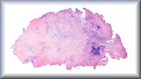
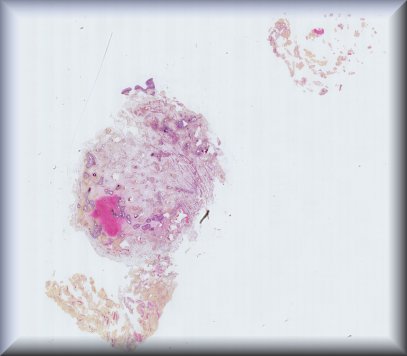
A 66-year-old male with progressive vision loss in the right eye and a history of pulmonary carcinoid.
Corneally displaced conjunctival melanoma with BRAF V600E mutation in a 48 year-old woman
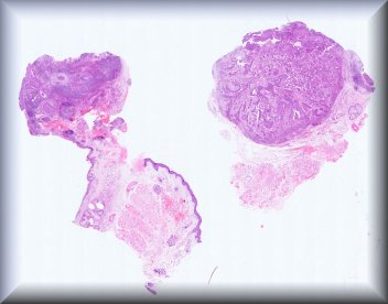
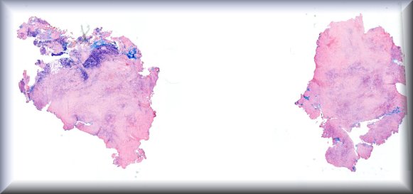
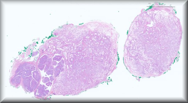
A 66-years-old man with blurry vision for one month in his left eye showed two choroidal tumors with different clinical and morphologic characteristics.
A 76-year-old woman presented with a long-standing history of left-sided epiphora, which since July 2025 has been progressively accompanied by nasal obstruction.
Jolique van Ipenburg (Nijmegen):
A 58-year old male patient presented with a saddle nose deformity and exophthalmos. PET and MRI revealed an extraconal orbital mass and soft tissue thickening alongside the nasal septum. Serum immuunglobulin G subclass 4 (IgG4) level was slightly elevated and anti-neutrophil cytoplasmatic antigen (ANCA) was negative.
7-year-old female patient. Right temporal conjunctival lesion that appeared approximately 2 years ago. Initially transparent and progressively enlarging. Lesion measuring 8 mm in greatest dimension. Clinically, a cystic lesion with a watery appearance and fluid content.
68 year-old male presented with chronic marked swelling of bilateral eyelids for more than a decade, despite bilateral upper lid blepharoplasty. Specimen is eyelid excision from blepharoplasty.
A 73-year-old man presented with epiphora and a medial canthal mass. MRI demonstrated a 2.5-cm lesion expanding the lacrimal sac with extension into the nasolacrimal duct. Excision of the lacrimal drainage apparatus was performed. Histology revealed an invasive adenosquamous carcinoma with a distinctive combination of features, including origin from a pre-existing papilloma, low-risk HPV-11, and ciliated glandular differentiation within the invasive component.
An 83-year-old man with a rapidly growing eyelid tumour is admitted for removal via Mohs surgery, without a prior biopsy.
55-year-old male with severe chronic ocular surface disease after toxic epidermal necrolysis, presenting with a right corneal opacity clinically suspicious for recurrent fungal keratitis
75-year-old female with B symptoms, including weight loss, presenting with multiple pigmented lesions of the eyelid and conjunctiva.
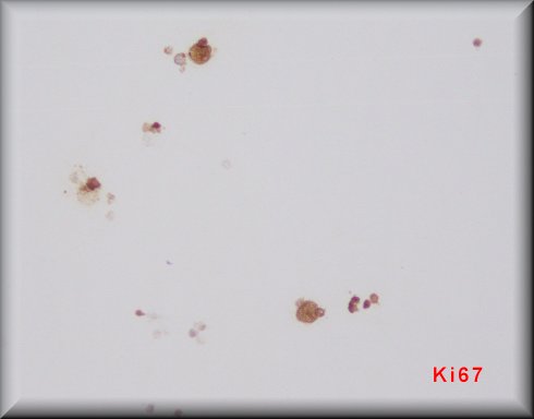
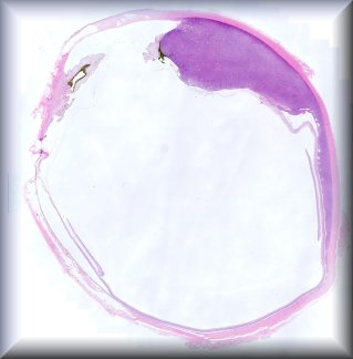
Patient with progressive visual deterioration and suspected intermediate uveitis without response to therapy; vitreous sample obtained for diagnostic evaluation.
32 year old male with large left intra and extraconal mass extending to lower lid. Biopsy of lesion.
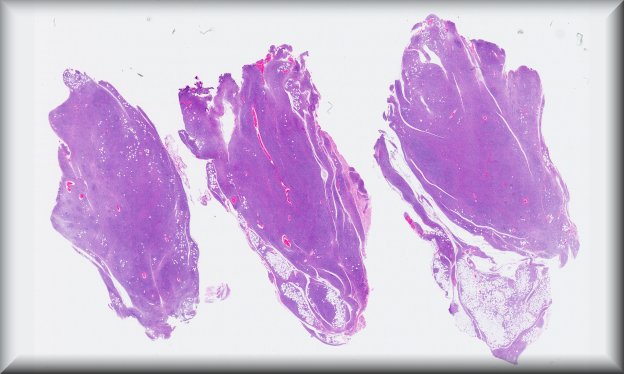
62 years-old male with a 2,5 x 2,0 cm lesion in the superior left eyelid rapidly evolving for nearly one month. The lesion was apparently centered in the eyelid rim, growing between the tarsus and the orbicularis oculi muscle. The patient underwent surgery in our hospital and we received the surgical specimen
Woman aged 40 years – excisional biopsy from the tumorous infiltrate of the left orbit (medial part).
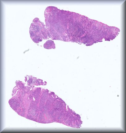
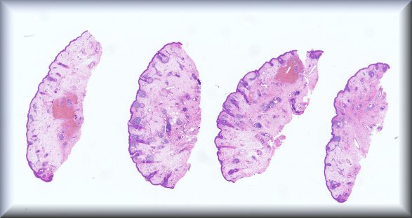
Enucleation for recurrence at the edge of the scar 2 years after brachytherapy for a choroidal and ciliary body melanoma clinically diagnosed at age 78 years.
A 60-year-old man with no significant past medical history was referred to the orbital unit for initial evaluation of left orbital swelling. His ophthalmologic history was notable for a prior episode of posterior scleritis. The patient provided a MRI study, with findings suggestive of an orbital inflammatory pseudotumor. The slide corresponds to a sample of the incisional biopsy of the left orbit.
Melike Pekmezci (San Francisco):
The patient is a 66-year-old man who presented a 1-year history of progressive eyefullness, irritation, dryness and intermittent lacrimation. Briefly, right eye showed periorbital fullness and resistance to retropulsion, but otherwise ophthalmic exam including vision, ocular surface, anterior chamber and retina were normal. normal vision, Imaging (MRI) Studies: CT scan at an outside hospital (March 2025) showed a right orbital mass. Follow-up CT scan at UCSF showed an infiltrative, enhancing mass involving the right lacrimal gland, with extension of abnormal enhancing tissue around the temporal and inferior aspect of the right anterior globe into the soft tissue of the left cheek.
Hardeep Singh Mudhar (Sheffield):
72-y-old female who presented 3 months after a right ptosis repair, with a right thickened eyelid mass, getting progressively larger. On palpation the tissue was firm. A biopsy was taken.
An 80-year old Caucasian male presenting with a slow growing right lateral canthal lesion query BCC. No change in vision. Eye examination otherwise normal. No previous significant ocular history. Known significant cardiovascular comorbidities.
A 26-year-old woman with a recurrent, massive retrobulbar tumor of the orbit
A 49-year-old patient presented with a conjunctival mass in the lower fornix of her left eye. She had suffered from a conjunctival carcinoma in situ 10 years earlier.
Rapidly evolving diplopia in a 14-yo boy. Excision of an upper orbital mass and biopsy of the supratroclear area.
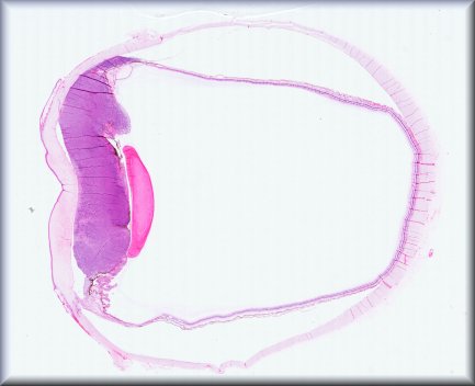
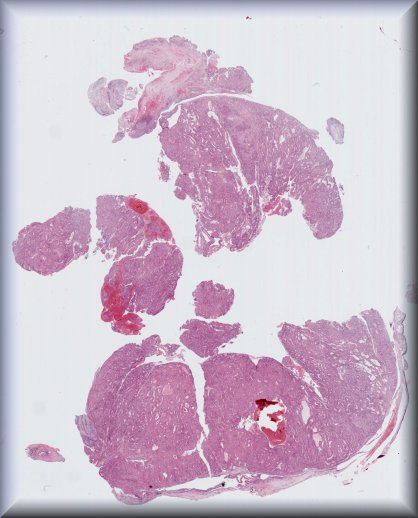
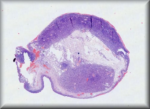
Man, 29 years-old. Enucleation of the left eyeball due to a round intraocular lesion on ultrasound; blind and painful eye
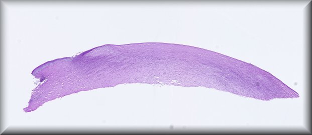
A 79-year-old man presented to the ophthalmology emergency department with a corneal foreign body at 5 o’clock and a pericorneal inflammatory reaction. The foreign body was removed and sent for analysis.